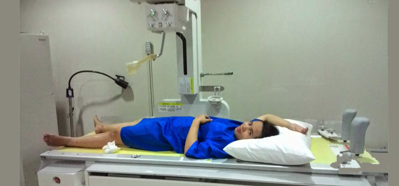
HYSTEROSALPINGOGRAPHY (HSG)

A blocked Fallopian tube or a growth in the uterus can significantly reduce chances for pregnancy. If the Fallopian tubes are blocked, the sperm can’t reach the egg to fertilize it.
What is Hysterosalpingography (HSG)?
A hysterosalpingogram, or HSG is an important test of female fertility potential. The HSG test is an X-Ray test usually done in the radiology department of a hospital or outpatient radiology facility.
- A Radiographic contrast (dye) is injected into the uterine cavity through the vagina and cervix
- First the uterine cavity fills with dye and since the uterus and the fallopian tubes are hooked together, the dye fills the tubes and spills into the abdominal cavity IF the tubes are Open.
Pictures are taken using a steady beam of X-ray as the dye passes through the uterus and fallopian tubes. The pictures can show problems such as an injury or abnormal structure of the uterus or fallopian tubes, or a blockage that would prevent an egg moving through a fallopian tube to the uterus. A blockage also could prevent the sperms from moving into a fallopian tube and fertilizing an egg.
Why is HSG Done?
A hysterosalpingogram is done to:
- Find a blocked Fallopian tube. An infection may cause severe scarring of the Fallopian tubes and block the tubes, preventing pregnancy. Occasionally the dye used during a hysterosalpingogram will push through and open a blocked tube.
- Find problems in the uterus, such as an abnormal shape or structure, an injury,polyps,fibroid, adhesion or a foreign object in the uterus. These problems could result in heavy menstrual bleeding and/or repeated miscarriage.
- See whether surgery to reverse a tubal litigation has been successful.
How is the Procedure Done?
The hysterosalpingogram study takes about 15-20 minutes to perform. The test is usually done in the radiology department of a hospital so there is additional time for the woman to register at the facility and fill out a questionnaire and answer questions regarding allergies to medication etc.
Details of Procedure as follows:
- The patient lies on the table on her back and brings her feet by the side just as in a normal speculum examination or transvaginal ultrasound
- The doctor places a speculum in the vagina and visualizes the cervix.
- Either a soft, thin catheter is placed through the cervical opening into the uterine cavity or an instrument called Allies forceps is placed on the cervix and then a narrow metal cannula is inserted through the cervical opening.
- Contrast is slowly injected through the cannula or catheter into the uterine cavity. An x-ray picture is taken as the uterine cavity is filling and then additional contrast is injected so that the tubes should fill and begin to spill into the abdominal cavity.
- More x-ray pictures are taken as this “fill and spill” occurs.
- The procedure is now complete. The instruments are removed from the cervix and vagina.
- The patient usually remains on the table for a few minutes to recover from any cramping caused by injection of the contrast.
The results of the test can be immediately available. The x-ray pictures can usually be reviewed with the patient several minutes after the procedure is done.
Does HSG have any side-effects Or Complications?
Complications commonly associated with a hysterosalpingogram include cramping and spotting.
Rarely HSG may be associated with infections, for which we routinely prescribe prophylactic antibiotics for three days. after the procedure
Results
| NORMAL: | Uterus looks normal in shape. Fallopian Tubes look Normal. Fallopian Tubes do not seem scarred or damaged. The dye flows into the uterus, then through the Fallopian Tubes and spills normally into the belly. |
|---|---|
| Uterus does not show any foreign objects (such as an IUD), tumors, or growths. | |
| ABNORMAL: | The dye does NOT flow through the Fallopian Tubes into the belly hence proving that the Tubes may be scarred, malformed OR blocked. Possible causes of blocked Fallopian tubes include Pelvic Inflammatory Disease, Endometriosis. |
| The dye may leak through the wall of the uterus, proving a tear or hole in the uterus. | |
| An abnormal uterus may show tissue (called a septum) that divides the uterus. | |
Growths, such polyps or fibroids may be present in the Uterus |
